
Consistent, documented microscopy figures.
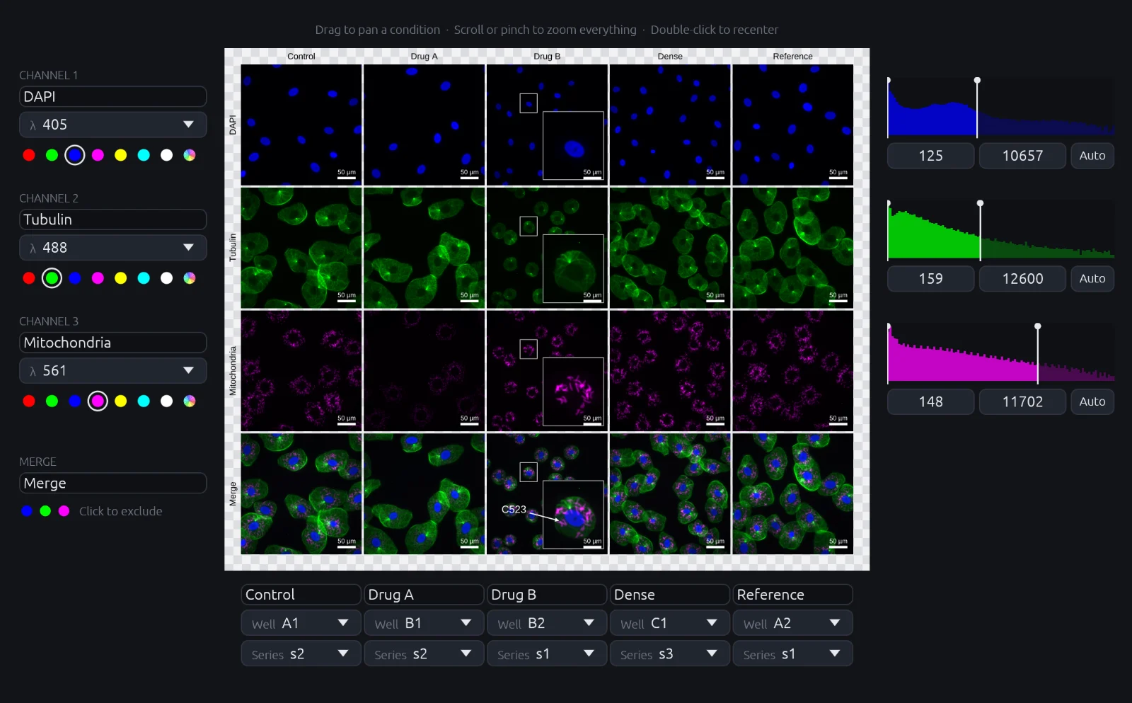
At the Journal of Cell Biology, a quarter of accepted papers had a figure sent back for image adjustments that broke the rules (Rossner). In cellernaS, each channel has one contrast setting for every condition, so panels can't be adjusted unevenly. The methods text is generated from those settings. A figure takes minutes, not an afternoon.
Download demo Express interest
macOS (Apple silicon, Intel). Not yet notarized: on first launch, open System Settings › Privacy & Security and click Open Anyway.
Fast, no slips
Drop in a folder. The grid builds itself from your file names: nothing to crop, align or rescale by hand.
Change it later
Adjust a range or swap a condition at any point. Every panel and the methods text update.
Reuse your settings
Save channels, colours and layout as a template. Apply it to the next experiment in one click.
Journal-ready output
TIFF/PNG at the journal's width and DPI, or PDF/SVG with editable labels.
Runs locally. Images never leave your computer.